3D Printed Models of Normal and Abnormal Fetal Hearts
Helena Dodziuk
hdodziuk@gmail.com
3D printing is widely used in industry but medicine is one of the main and most varied areas of its applications. These are not limited to inexpensive and personalized prostheses and implants but also used for printing personalized (for surgery on infants and fetuses [1]) and to manufacture inexpensive medical devices, medications (the first one approved by FDA) and models for surgeons allowing them to plan surgical procedures. Cell printing is also used in sophisticated embryonic stem cells printouts and organ models for drug testing, Dr. Marcin Wiecheć with his co-workers at Jagiellonian University and the company Grid, both located in Cracow, Poland, launched the project “3D cardiac models for prenatal diagnostics.” The project’s aim is to improve prenatal detection of cardiac anomalies by producing 3D printouts presenting normal fetal heart and about forty variations of cardiac anomalies. The layered structure of 3D printed cardiac models illustrates prenatal ultrasound images and allows for better understanding of the geometry of normal and abnormal fetal hearts by physicians involved in prenatal diagnostics.
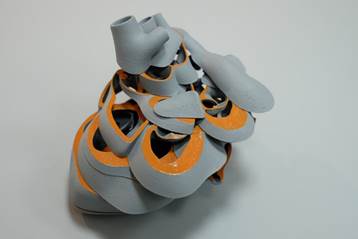
Fig. 1. A 3D model of a normal fetal heart.
Prenatal sonography is one of the major screening tools used during pregnancy to check the fetal development. Compared to other methods of medical imaging techniques, sonography has important advantages such as real-time imaging capability. Ultrasound scanners are easily movable; the examination is often substantially lower in cost then other imaging techniques.. From the technical point of view, sonography is based on emitting and receiving an ultrasound beam. It does not involve any harmful ionizing radiation. The drawbacks of sonography include imaging difficulties for tissues located behind bones and airways, penetration problems in high BMI patients, and its dependency on an operator. Operators are trained in obtaining the best available visualization conditions through so-called acoustic windows. Nevertheless, mage interpretation skills require a steep learning curve.
Fetal heart can be seen during a scan at the 5th week of gestation, but its structure can be evaluated at the earliest by about 12 week when the fetus attains approximately 5 mm in length. At this time, all major components of the heart including atria, ventricles and arteries are visible and congenital heart defects can be identified allowing physicians to monitor the fetal well-being and, eventually, prepare for future postnatal surgery. Some anomalies, like the bicuspid instead of tricuspid aortic valve are less significant, others, such as a complete atrio-ventricular canal, CAVC, will require surgery in the postnatal period. The prenatal diagnosis is essential for future treatment. Dr. Wiecheć created the models that help doctors to understand complicated spatial relationships among organs forming the fetal heart. The models may be used as well when counseling parents.
The development of models was not easy. Initially, the participants experienced typical problems related to cooperation of specialists from different areas. Doctors found it difficult to visualize the internal structure of the heart. Designers from Grid specializing in industrial and interior design had no experience in anatomy, to say nothing about prenatal anatomy, They began by reviewing 3D and 2D ultrasound images and transferring them into 3D Computer Graphics volume images. At early stages of the project development, they even made plasticine models. At the end, an experienced cardiac surgeon, Dr. Jacek Kolcz, reviewed all 3D models. In order to be used for teaching purposes, the models can be disassembled and reassembled into representative layers corresponding to prenatal cardiac sonographic views by using 60 magnets embedded in the plastic structure.
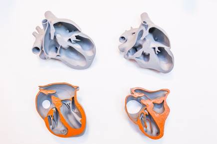
Fig. 2. Two cardiac 3D models opened at the level of four-chamber view (left, normal fetal heart; right, the heart exhibiting hypoplastic left heart syndrome based on aortic atresia.
The models help in diagnosis of heart malformations and then in explaining to parents the need for heart surgery for their child. They became an officially endorsed teaching aid and are used by Malopolskie Ultrasonographic Workshops. Mass production of models is being planned. Dr. Fred Ushakov, Fetal Medicine Specialist, University College London Hospital, UK, and Ambassador of International Society of Ultrasound in Obstetrics and Gynaecology, ISUOG, expressed the following opinion: “I consider the early diagnosis of fetal heart malformations to be the most important current task for Fetal Medicine. I am delighted with the brilliant idea of Dr. Marcin Wiechec to create 3D printed models of normal and malformed fetal hearts and their elegant technical implementation. Those models are practically perfect in their illustration of complex three-dimensional anatomy of the heart and represent an unique tool for all professionals dealing with prenatal diagnosis of congenital heart disease and cardiac surgery.”
Consider an example of one of the most difficult to diagnose before birth defects: D-Transposition of Great Arteries (d-TGA). Because of the difficulty of understanding the different geometry of a fetal heart affected by this condition from that of a healthy one, the general detection rate of d-TGA is only 40%. 3D models will definitely help doctors in making the diagnosis. If this anomaly is detected prenatally, optimal conditions for delivery at the tertiary center will be arranged and required surgery within 2 weeks after delivery will increase the chances of survival for babies with d-TGA.

Fig. 3. Analyzing models during one of the ultrasound workshops in Cracow, Poland.
Gruijthuijsen, C., Rosa, B., Snyers, H., Engels, A., Vercauteren, T., Deprest, J., Ourselin, S., Reynaerts, D., Vander Poorten, E. (2015). Prototyping Novel Instruments for Fetal Surgery through Virtual Reality Simulation and 3D Printing. (Proceedings of the 5th Joint Workshop on New Technologies for Computer/Robot Assisted Surgery. Joint Workshop on New Technologies for Computer/Robot Assisted Surgery (pp. 98-101))